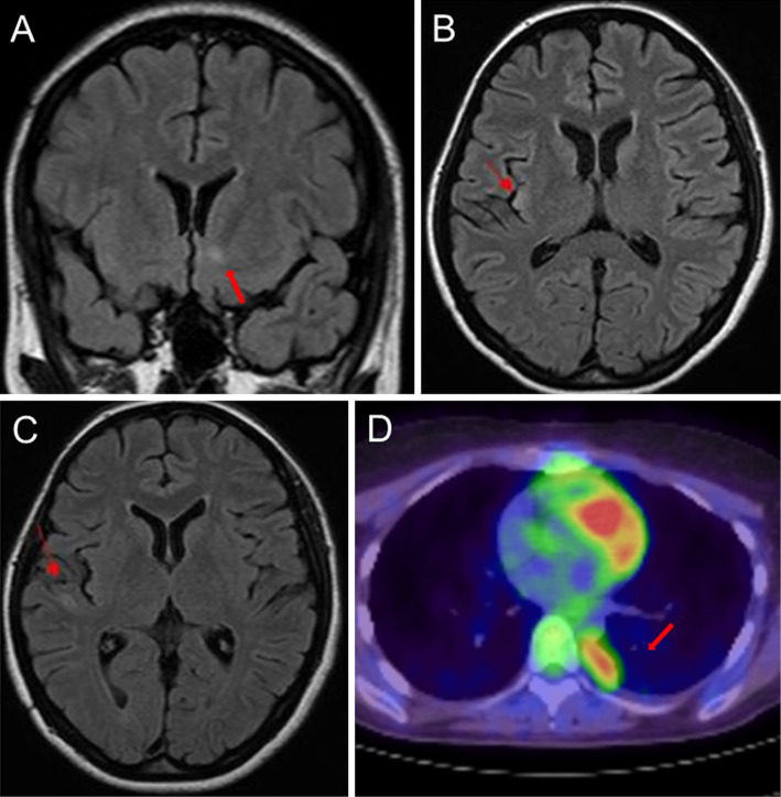
Figure.
Brain magnetic resonance imaging and positron emission tomography/computed tomography (PET/CT) of the thorax. (A) On admission, a hyperintense lesion was identified in the lower region of the left caudate nucleus by fluid-attenuated inversion recovery (FLAIR) imaging. (B) On day 14 of hospitalization, a new lesion was identified in the right insula by FLAIR imaging. (C) On day 54 of hospitalization, a new lesion was identified in the right temporal lobe by FLAIR imaging. (D) Preoperative chest PET/CT images showed a 1.2-cm mass at the base of the right lung, which indicated thymoma recurrence.

