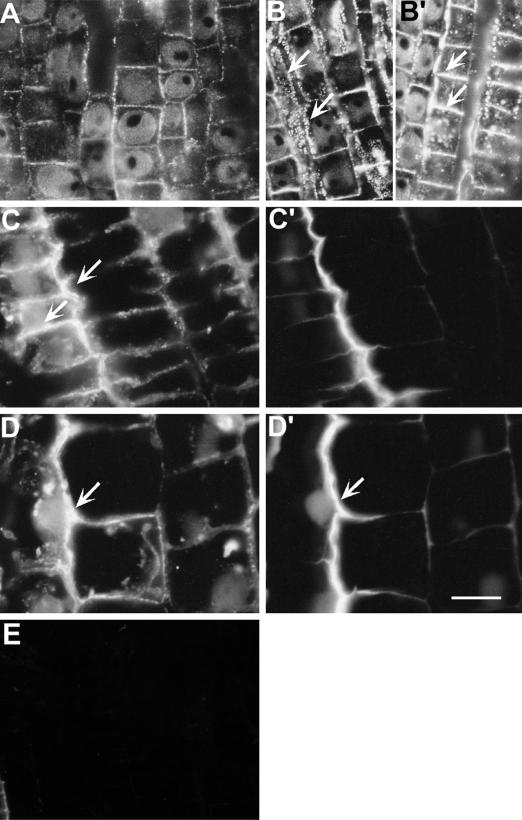
Figure 8.
Effect of Al (20 μm, 3 h) on the expression of calreticulin probed with polyclonal antibody (immunofluorescence technique) and its colocalization with Al-induced callose in wheat cv Scout 66 root apex. Control image (A) is showing basal fluorescence of calreticulin preferentially lining the cortex cell pit-field peripheries as fine dots. Increased fluorescence in the same pit-field regions was apparent after Al treatments (B, B′, C, and D). Arrows in B and B′ showing cortical cells with enhanced intensity both from the longitudinal and transverse regions of cell peripheries. Similar expression was found at epidermal cells (arrows in C) and the same regions were characterized with the enhanced callose (C′). This pattern of colocalization continued all along the root apex as evidenced by the expression at the middle part of transition region epidermal cells (D) and the callose version (D′) of the same frame (arrows). Note that the callose is not appearing as fine dots since this region of section is not paradermal (see text for details). Sections labeled without the primary antibody showed no fluorescence signal (E). Representative images of at least two independent experiments comprising four to six individual root apices. Scale bar represents 18 μm.

