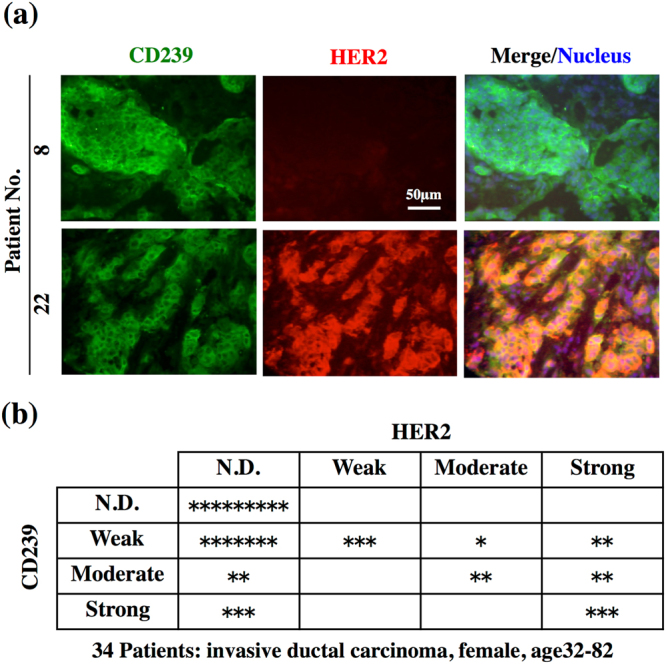
Figure 1.

Expression of CD239 and HER2 in breast cancer tissues. (a) Frozen tissue sections were stained with antibodies against CD239 (green, left panels) and HER2 (red, centre panels). Merged images of CD239 and HER2 staining are shown in the right panels. Nuclei were counterstained with Hoechst 33258 (blue). CD239 is highly expressed in tissues of patients 8 and 22. (b) Summary of CD239 and HER2 expression in the breast cancer tissues tested. The scale of expression is described in Methods. N.D., not detected. *, a classified tissue.
