
Figure 4.
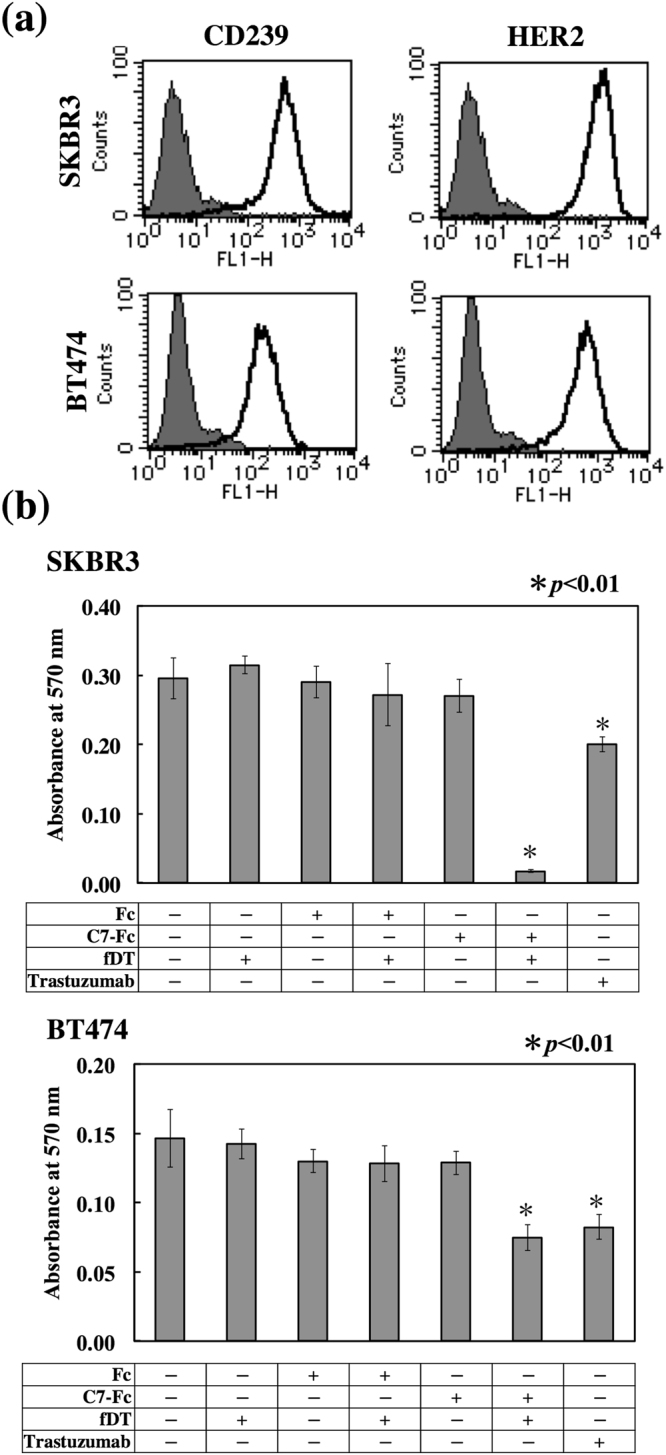
Cytotoxicity of fDT-bound recombinant antibody on CD239-highly positive breast cancer cells. (a) Flow cytometric analysis of CD239 and HER2 expression in SKBR3 and BT474 cells. Solid lines show C7-Fc and trastuzumab antibodies, and grey fill indicates the control Fc recombinant protein. (b) Cytotoxicity of fDT-bound C7-Fc. The recombinant proteins (C7-Fc and Fc) were combined with the fragment of diphtheria toxin (fDT). SKBR3 and BT474 cells were cultured in the presence of the recombinant proteins or fDT-bound recombinant proteins. After 4 days in culture, the number of cells was determined.