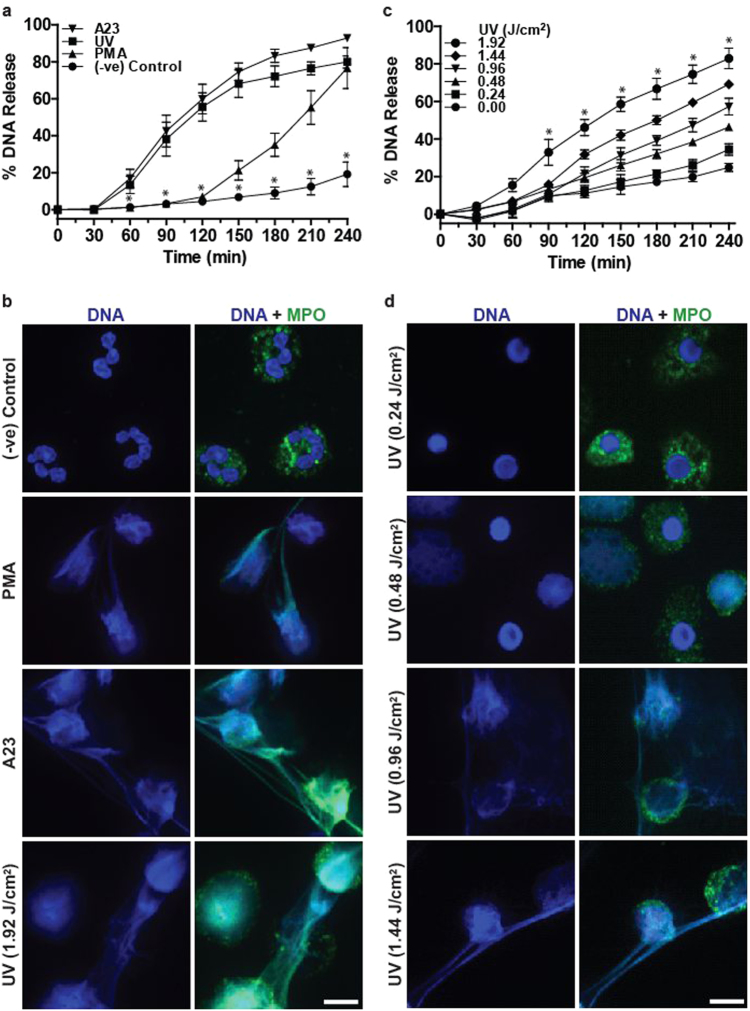
Fig. 1. UV induces rapid NETosis and increasing UV dose increases NETosis.
a DNA release from neutrophils following media (−ve control), PMA (25 nM) and A23 (4 µM) treatment or UV irradiation (1.92 J/cm2) was measured using the SYTOX Green plate reader assay. UV-induced NETosis follows the kinetics similar to that of A23-induced NETosis (n = 3; error bars represent SEM; A23, A23187; *p < 0.05). b Neutrophils were treated with PMA (25 nM), A23 (4 µM) or UV (1.92 J/cm2) and incubated for 240 min. Cells were stained for DNA (DAPI, blue) and MPO (green). Immunofluorescence imaging shows that MPO colocalises to DNA, hence 1.92 J/cm2 UV induces NETosis. Images are representative of three independent experiments. Scale bar, 10 μm. See low-magnification images in Fig. S2. c DNA release following treatment with increasing dose of UV was measured using the SYTOX Green plate reader assay (n = 3; error bars represent SEM; *p < 0.05). Increasing dose of UV results in increased NETosis. d Neutrophils were treated with varying doses of UV and incubated for 240 min. Cells were stained for DNA (DAPI, blue) and MPO (green). Confocal fluorescence imaging shows that UV light exposure results in NETosis at higher doses and apoptosis at lower doses. Images are representative of three independent experiments. Scale bar, 10 μm. See low-magnification images in Fig. S3

