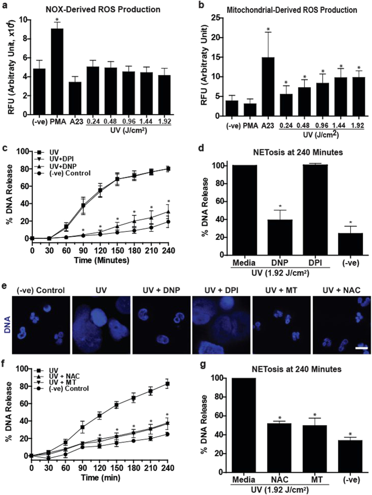
Fig. 3. Mitochondrial but not NOX-mediated ROS production is involved in UV-induced NETosis.
a NOX-derived ROS production was measured using the DHR123 plate reader assay. Cells were treated with PMA (25 nM), A23 (4 µM) or increasing doses of UV, and the fluorescence signals were recorded. None of the doses of UV induce NOX-derived ROS, similar to A23, except PMA that induces a significant amount of NOX-mediated ROS production (n = 3; error bars represent SEM; A23, A23187; *p < 0.05 for comparing with the negative control). b Mitochondrial ROS production was measured using the MitoSOX plate reader assay. Higher doses of UV induce mitochondrial-derived ROS, similar to A23 (4 µM), but unlike PMA (25 nM) that does not induce a significant amount of mitochondrial ROS production (n = 3; error bars represent SEM; A23, A23187; *p < 0.05 for comparing with the negative control). c, d DNA release following UV (1.92 J/cm2) treatment was measured using the SYTOX Green plate reader assay (n = 3). Cells were preincubated either with DPI (1 µM) or DNP (750 µM) for 1 h prior to UV treatment. DNP, but not DPI, significantly inhibits UV-induced NETosis. e Neutrophils were incubated with NAC (3 mM), MitoTempo (100 µM), DPI (1 µM) or DNP (750 µM) for 1 h and then treated with UV (1.92 J/cm2) and incubated for 240 min without SYTOX green. Cells were stained for DNA (DAPI, blue). Fluorescence imaging shows that NAC, MitoTempo and DNP, but not DPI, inhibit UV-induced NETosis. Images are representative of three independent experiments (scale bar, 10 μm). f, g DNA release following UV (1.92 J/cm2) treatment was measured using the SYTOX Green plate reader assay (n = 3). Cells were incubated with NAC (3 mM) or MitoTempo (100 µM) for 1 h prior to UV treatment. NAC and MitoTempo significantly inhibit UV-induced NETosis. Error bars on graphs represent SEM. MT, MitoTempo; *p < 0.05 for comparing UV treatment alone to UV treatment with inhibitors

