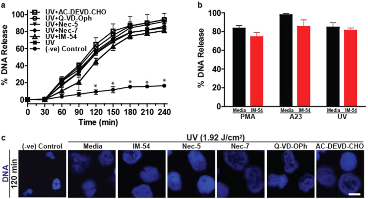
Fig. 7. UV-induced NETosis differs from UV-induced apoptosis, necrosis and necroptosis.
a, b DNA release following UV (1.92 J/cm2) treatment was measured using the SYTOX Green plate reader assay (n = 3). Cells were incubated with IM-54 (10 µM), necrostatin-5 (10 µM), necrostatin-7 (10 µM), QD-VD-Oph (10 µM) or AC-DEVD-CHO (10 µM) for 1 h prior to UV treatment. None of the inhibitors significantly inhibit UV-induced NETosis. c Experiments were conducted as above, except that SYTOX Green was omitted from the assay, and samples were incubated for 120 min. Cells were stained for DNA (DAPI; blue). Fluorescence imaging confirms that none of the inhibitors suppress UV-induced NETosis (scale bar, 10 μm). Images are representative of three independent experiments. Error bars represent SEM; *p < 0.05 for comparing UV treatment alone to UV treatment with inhibitors. See low-magnification images in Fig. S6

