Figure 1.
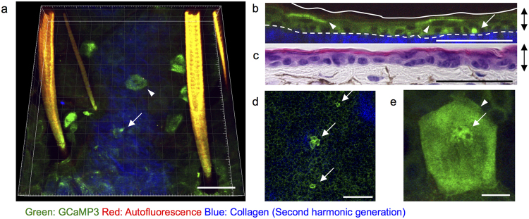
Physiological state of [Ca2+]i in the epidermis. Representative two-photon images of the ear skin of mice systemically expressing GCaMP3 (GCaMP3 mice). (a) Three-dimensional view, (b) Vertical view showing cells with elevated fluorescence of GCaMP3 (GCaMP3high cells). Arrow and arrowhead: GCaMP3high cells in the basal and granular layers, respectively. Green: GCaMP3; red: autofluorescence; blue: second-harmonic generation from dermal collagen. Dashed line in (b): dermo-epidermal junction. (c) Hematoxylin and eosin staining of the ear skin section. Double-headed arrows: epithelial layers. (d) Horizontal view of the basal layer. Arrows: GCaMP3high basal cells. (e) Maximum intensity view of the granular layer. Arrowhead: a GCaMP3high granular cell. Arrow: nucleus. Scale bars: 50 μm (a–d) and 10 μm (e). [Ca2+]i: concentration of calcium ions in the cytoplasm. Results are representative of at least three independent experiments.

