
Figure 7.
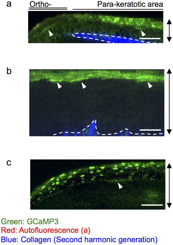
Physiological state of [Ca2+]i in the parakeratotic epidermis and mucous epithelia. Representative two-photon images of GCaMP3 mice (a,b) or GCaMP3/tdTomato (c), in a vertical view. (a) In vivo observation of the tail skin. (b,c) Ex vivo observation of the mucous epithelia of the (b) vagina and (c) oral cavity. The contrast in the fluorescence of GCaMP3 was corrected using the fluorescence signal of tdTomato, which reflects the concentration of fluorescent proteins in the cytoplasm. Arrowheads: cells with elevated fluorescence of GCaMP3 beneath the cornified layer. Dashed lines: dermo-epidermal junction or lower edge of the mucous epithelia. Green: GCaMP3; red: autofluorescence (a); blue: second-harmonic generation from dermal collagen. Double-headed arrows: epithelial layers. Scale bars: 50 μm. Results are representative of at least three independent experiments.