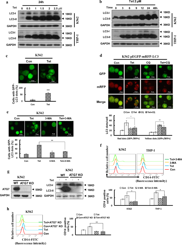
Fig. 3. Autophagy played an important role in tetrandrine-induced differentiation.
The bars indicate the S.D. (a) K562 and THP-1 cells were treated with the indicated concentration of tetrandrine for 24 h or (b) with 2 μM tetrandrine for the indicated durations. Western blot analysis of LC3 levels. GAPDH was used as a loading control. (c) K562 cells were transfected with the GFP-LC3 plasmid for 24 h, subsequently treated with DMSO or 2 μM tetrandrine for 24 h and observed by fluorescence microscopy. Representative experiments are shown to indicate the cellular localization patterns of the GFP-LC3 fusion protein (magnification ×400), and percentage of cells with GFP-LC3 puncta were used to quantify the percentage of autophagic cells. n = 3, ***p < 0.001. (d) After a 1 h pretreatment with 10 μM CQ and 48 h of subsequent treatment with DMSO or 2 μM tetrandrine, fluorescence microscopy detected green and red fluorescent spots in K562 cells transfected with the ptfLC3 plasmid and percentage of cells with red dots were used to quantify the autophagic flux, while yellow dots (the overlay of green and red fluorescence) were increased if the process of autophagosomes fusing with lysosomes was inhibited. n = 3, *p < 0.05, **p < 0.01. (e) GFP-LC3-transfected cells were pretreated with or without 1.5 mM 3-MA for 1 h. The cells were then exposed to DMSO or 2 μM tetrandrine for 24 h, and the localization of GFP-LC3 was observed using a fluorescent microscope (magnification ×400). n = 3, **p < 0.01. (f) CD14 antigen expression was analyzed by flow cytometry after treatment for 4 days with or without pretreatment with 1.5 mM 3-MA for 1 h, and a statistic analysis for three experiments in the bottom. *p < 0.05. (g) The expression of ATG7 and LC3-II proteins in K562 ATG7 KO cells were assessed via western blot, and K562 WT cells were used as a positive control. (h) CD14 expression was determined by flow cytometry in K562 ATG7 and K562 WT cells after treatment for 4 days, and a statistic analysis for three experiments in the right. **p < 0.01. WT wild type

