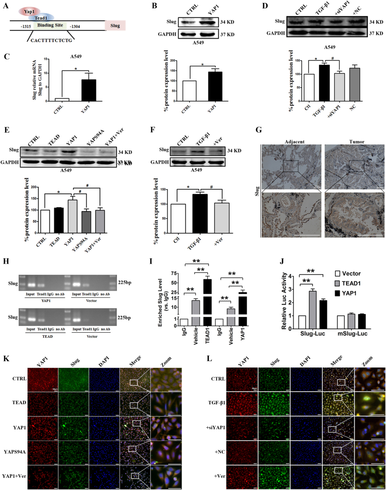
Fig. 6. Slug is regulated by the co-transcriptional complex YAP1/TEAD in the EMT program of A549 cells.
a The gene sequence analysis shows a putative TEAD binding site in the promoter of Slug. Western blot (b) and real-time RT-PCR (c) assays show that YAP1 overexpression upregulates the protein and mRNA levels of Slug in A549 cells. GAPDH was used as an internal control. Slug mRNA levels were normalized to GAPDH. *P < 0.05 vs. CTRL. d–f. Western blotting was used to analyze the expression of Slug in A549 cells. GAPDH was used as an internal control. d YAP1 silencing decreases Slug protein levels in A549 cells. *P < 0.05 vs. CTRL; #P < 0.05 vs. TGF-β1. e Verteporfin inhibits Slug expression, and YAPS94A has no effect on Slug expression in A549 cells. *P < 0.05 vs. CTRL; #P < 0.05 vs. YAP1. f Verteporfin reverses the upregulation of Slug via TGF-β1 in A549 cells. *P < 0.05 vs. CTRL; #P < 0.05 vs. TGF-β1. g Representative images of the immunohistochemical (IHC) staining of Slug in human NSCLC tissues and matched adjacent tissues show that a significant increase in YAP1 staining is found in human NSCLC tissues. n = 10. qPCR (h) and chromatin immunoprecipitation (ChIP) (i) assays demonstrate the physical interaction between TEAD and the promoter region of Slug. *P < 0.05 vs. IgG. j Luciferase assays confirm that TEAD can active the transcription of Slug-luc but not mSlug-luc. *P < 0.05 vs. Vector. k, l Immunofluorescence assays show the staining intensities of YAP1 and Slug in A549 cells. YAP1 is stained red, Slug is stained green, and the nuclei are stained blue. The scale bars indicate 50 µm. The experiments were performed three times, and the data are presented as the mean ± SEM. n = 4–8

