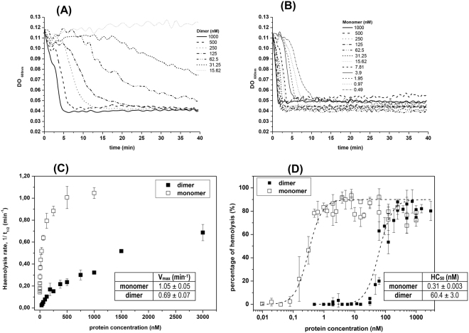
Figure 4.
Hemolytic activity of monomer and dimer StI W111C forms. Time course of erythrocyte lysis of homodimer (A) and monomer (B) at different concentrations by measuring the decrease in the turbidity (DO600 nm) of an erythrocyte suspension. (C) dependence of the haemolytic rate respect to monomer and dimer concentrations. The kinetic parameters t1/2 (time when the initial DO600 nm value of the erythrocyte suspension is reduced to the half) was estimated from curves on panels A and B. Rate of hemolysis is expressed as the reciprocal of the half-time of hemolysis (t1/2). (D) The percentage of hemolysis of dimer (without 2-ME) and monomer (dimer incubate with 0.1 M of 2-ME for 24 h) was determined for various doses of the toxin. The HC50 (concentration where 50% of HRBC are lysed after 15 min) of dimer (solid squares) and monomer (open squares) calculated from the best sigmoidal fit (dash lines) are shown in the inserted table. In panels C and D are shown the means and standard errors from 3–5 independent experiments.

