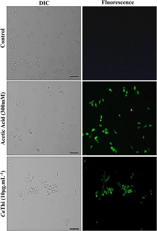
Figure 2. Activity of caspase in C. tropicalis cells after 24 h of incubation with 10 μg.ml−1 of CaThi.
Control cells and cells treated with CaThi were incubated with the FITC-VAD-FMK probe and analyzed by fluorescence microscopy. Green fluorescence indicates positive staining for caspase activity. Bars = 10 μm.

