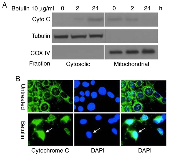
Figure 5.

Release of cytochrome c in apoptosis induced by betulin in HCT116 cells. Cells were treated with betulin (10 µg/ml) for 0, 2 and 24 h. (A) Cytosolic and mitochondrial fractions isolated from cells. The distribution of cytochrome c was analyzed with western blotting. The control groups for fractionation were α-tubulin and cytochrome oxidase subunit IV. (B) Immunofluorescent staining of HCT116 cells with cytochrome c (green) and DAPI (blue). Cyto C, cytochrome c; COX IV, cytochrome oxidase IV.
