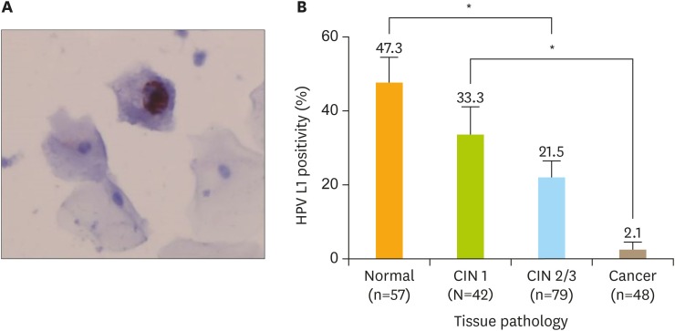
Fig. 1.
HPV L1 immunoreactivity in HPV16-positive cervical cytology. (A) Positivity of L1 capsid protein in a LSIL; note the presence of strong nuclear staining. The red nucleus represents L1 positivity (original magnification, ×400). B) HPV L1 positivity by cervical lesion.
CIN, cervical intraepithelial neoplasia; HPV, human papillomavirus; LSIL, low-grade squamous intraepithelial lesion.
*Asterisks indicate cervical lesions with significantly different percentages of L1 positivity.

