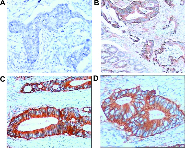
Figure 2.
Analysis of C-MET expression by immunohistochemistry in colorectal carcinomas. C-MET expression was localized in the membrane and its expression was observed predominantly in cancer cells. (A) Negative C-MET staining in a cancerous tissue sample (magnification, ×100). (B) Positive C-MET staining in tumor cells (upper), with negative or weak staining in adjacent epithelial cells (lower) (magnification, ×100). (C) Strong C-MET staining in tumor nests (magnification, ×100). (D) Positive membrane staining, as observed in the majority of tumor cells (magnification, ×200). C-MET, c-mesenchymal epithelial transition factor.

