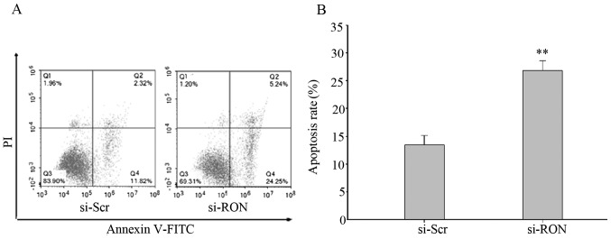
Figure 4.
Silencing RON induced apoptosis in 5637 cells. Flow cytometry was used to detect the proportion of cells in early and late apoptosis following transfection with si-RON, and compared with si-Scr. (A) Cells were transfected with si-RON or si-Scr for 48 h, collected, stained with Annexin V and PI and then analyzed by flow cytometry. (B) Quantitative results were obtained using Annexin V/PI staining. Data are derived from three independent experiments and are expressed as the mean ± standard deviation. **P<0.01, compared with si-Scr group. FITC, fluorescein isothiocyanate; RON, macrophage stimulating 1 receptor; si-Scr, scramble small interfering RNA; si-RON, human RON-targeting small interfering RNA; PI, propidium iodide.

