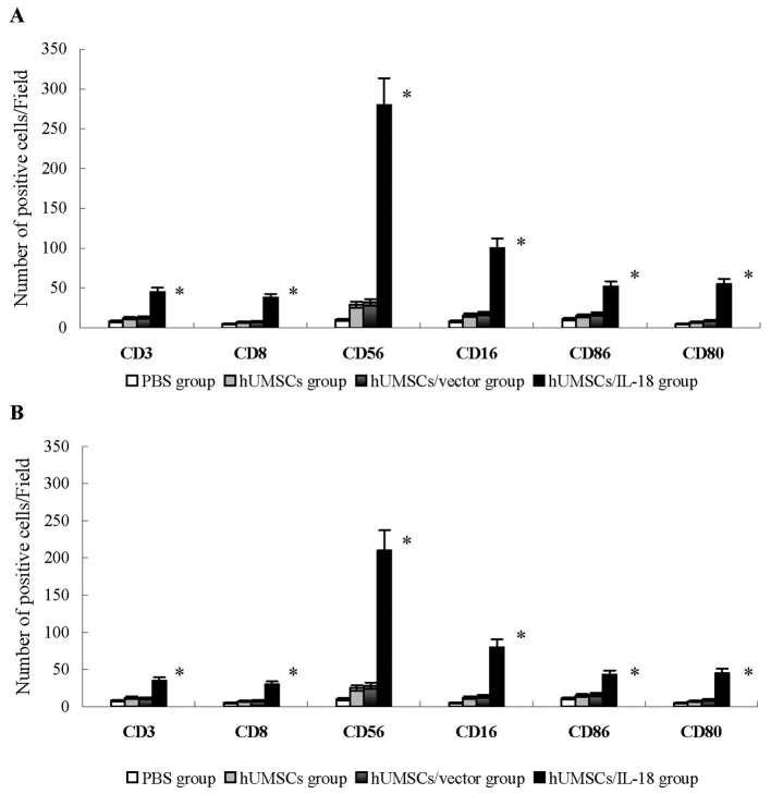
Figure 3.
Analysis of lymphocyte infiltration into tumor tissues. Tumor tissues were snap-frozen, and 4-µm thick sections were prepared, then stained with fluorescently labeled antibodies. The number of CD3+ and CD8+ T cells, and CD16+, CD56+, CD80+ and CD86+ NK cells were quantified in 4 sections randomly selected for each group. (A) In the early-effect study, the proportions of CD3+ and CD8+ T cells, and CD16+, CD56+, CD80+ and CD86+ NK cells in the hUMSC/IL-18 group were significantly increased compared with the other groups. (B) In the late-effect study, the proportions of CD3+ and CD8+ T cells, and CD16+, CD56+, CD80+ and CD86+ NK cells in the hUMSC/IL-18 group were significantly increased compared with those in the other groups, but were decreased compared with those in the hUMSC/IL-18 group in the early-effect study (P=0.039). *P<0.05 vs. all other groups. CD, cluster of differentiation; hUMSCs, human mesenchymal stem cells derived from umbilical cord; IL-18, interleukin 18; NK, natural killer.

