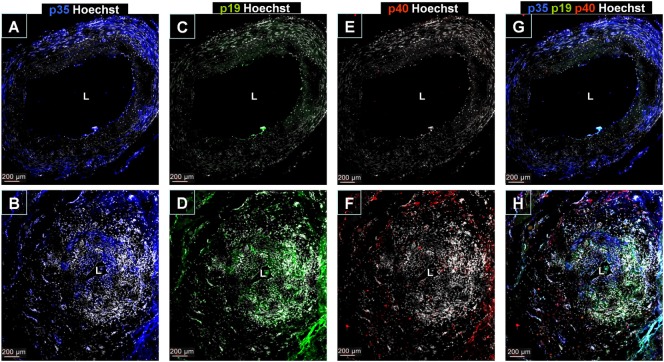
Figure 2.
Immunofluorescence detection of IL-12/23p40, IL-12p35, and IL-23p19 subunit expression in temporal artery lesions. (A) IL-12p35 staining (blue) in a temporal artery from a control individual showing almost selective expression in the media layer. Nuclei were stained with Hoechst (white). (B) IL-12p35 expression in GCA-involved temporal artery section, predominantly in inflammatory infiltrates. (C,D) Negative IL-23p19 immunostaining (green) in sections of normal temporal arteries (C) and intense expression in GCA samples (D) where IL-23p19 expression can be observed in all arterial layers especially in the most inflamed areas. (E) Lack of IL-12/23p40 immunostaining in a temporal artery from a control. (F) Detection of IL-12/23p40 expression (red) in a GCA-involved temporal artery section predominantly in the adventitial layer. (G,H) IL-12p35 (blue), IL-23p19 (green) and IL-12/23p40 (red) staining merge in a temporal artery section from a control and from a GCA patient, respectively. Pictures are representative of four arteries from four GCA patients and two controls, and at least three sections per sample were evaluated.

