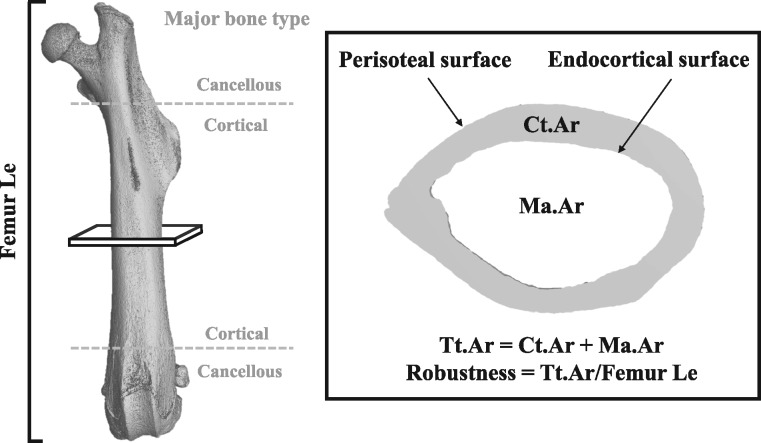
Figure 1:
major bone types and morphological parameters assessed by nano-computed tomography (nanoCT) in our exposure study. The shaft of the long bone (between the gray dotted lines) is primarily composed of cortical bone, while the distal ends contain more cancellous bone. Cross-sectional bone assessments (inset) in the mid-diaphyseal region of the femur included: Ct.Ar, cortical area (gray shaded region); Ma.Ar, marrow area (inner white region); Tt.Ar, total cross-sectional area (gray + inner white regions); Femur Le, femur length.

