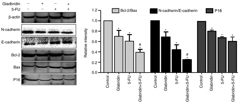
Figure 6.
Protein expression of MKN-45 cells was determined by western blotting in the presence or absence of glabridin and 5-FU. E-cadherin and Bax expression was significantly increased in the presence of glabridin or 5-FU, and reached a maximum in MKN-45 cells in the presence of glabridin combined with 5-FU. N-cadherin, Bcl-2 and p16 expression was significantly decreased in the presence of glabridin or 5-FU, and reached the lowest level in MKN-45 cells treated with glabridin combined with 5-FU. β-actin was used as a loading control. Data was obtained from a minimum of three independent experiments. *P<0.05 (vs. control group) or #P<0.05 (glabridin+5-FU vs. 5-FU) was considered to indicate a statistically significant difference. The molecular weights of E-cadherin and N-cadherin were 97 and 140 KDa, respectively; thus, the molecular weight was so large that the exposure time was extended. Therefore, the background of the E- and N-cadherin bands appears granulated and different from the background of the other bands. The same membrane was used. 5-FU, fluorouracil; Bcl-2, apoptosis regulator Bcl-2; Bax, apoptosis regulator BAX.

