Figure 1.
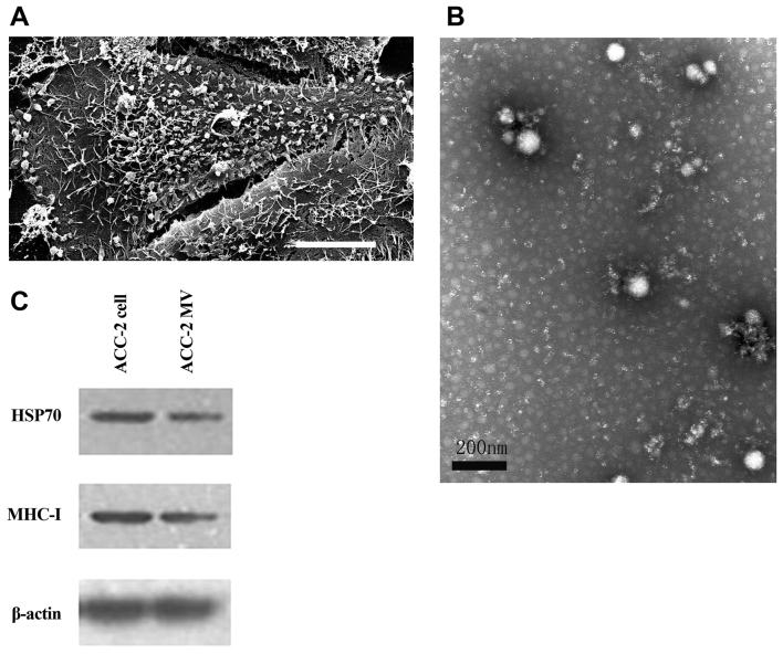
Characterization and identification of ACC-2 cell-derived MVs. (A) The scanning electron micrograph revealed that an ACC-2 cell was covered with MV-like structures varying in size from 30–500 nm. Scale bar, 10 µm. (B) ACC-2 MVs were isolated by differential centrifugations from medium supernatants and the pellets were examined by TEM. TEM image of the ACC-2 MVs depicting rounded structures with a size of 30–100 nm. Scale bar, 200 nm. (C) HSP70 and MHC-I protein levels in ACC-2 cells and their microvesicles were detected using western blot analysis. The β-actin loading control is also shown (n=3). MV, microvesicle; TEM, transmission electron microscopy; MHC-I, major histocompatibility complex class I; HSP70, heat shock protein 70.

