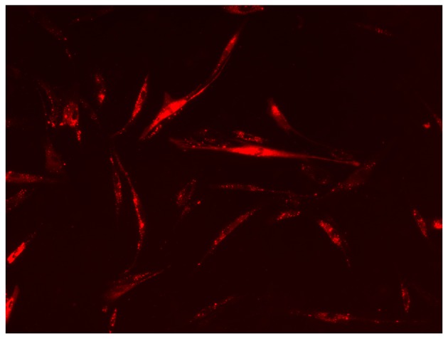
Figure 3.

ACC-2 MVs are internalized into RSC96 cells. The ACC-2 MVs labelled with the membrane dye DiI (red) or MV-free supernatant were added to RSC96 cells in culture. The cells were then observed using a fluorescence microscope at magnification, ×100. The image depicts red fluorescent-positive RSC96 cells. MV, microvesicle.
