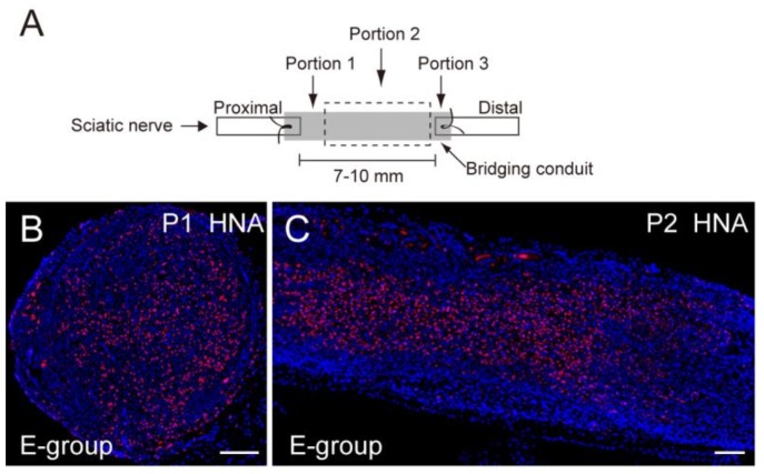
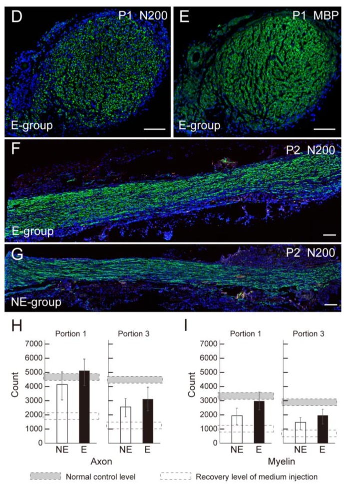
Figure 1.
Extraction of histological sections and comparison of the number of axons and myelin in the conduit. (A) Histological sections were obtained from three portions; Portions 1 and 3 are cross-sections, and Portion 2 is a longitudinal section; (B,C) Typical engraftment of human Sk-34 cells stained with HNA (human nuclear antigen, red) in cross- (B) and longitudinal (C) sections; (D,E) Typical staining of axons (D, N200, green), and typical staining of myelin (E, MBP; myelin basic protein, green) E-group; (F,G) Similarly, typical staining of axons (green) in longitudinal sections (Portion 2) both in E-group (F) and NE-group (G); Axonal regeneration is apparent through the conduit in both groups. Blue staining is nuclear staining of 4′,6-diamidino-2-phenylindole (DAPI). P1 and P2 correspond to Portions 1 and 2, respectively. Bars in B-E = 100 μm; (H) Axon numerical recovery in Portions 1 and 3; (I) Myelin numerical recovery in Portions 1 and 3. The E group consistently showed greater recovery of axon and myelin in the conduit than the NE group. NE = non-exercise group, E = exercise group. Gray dotted square shows the mean number of axons in the same portion of a normal control sciatic nerve based on our pooled data. An open dotted square shows the recovery level of the case of conduit + non-cell media injection derived from our pooled data.


