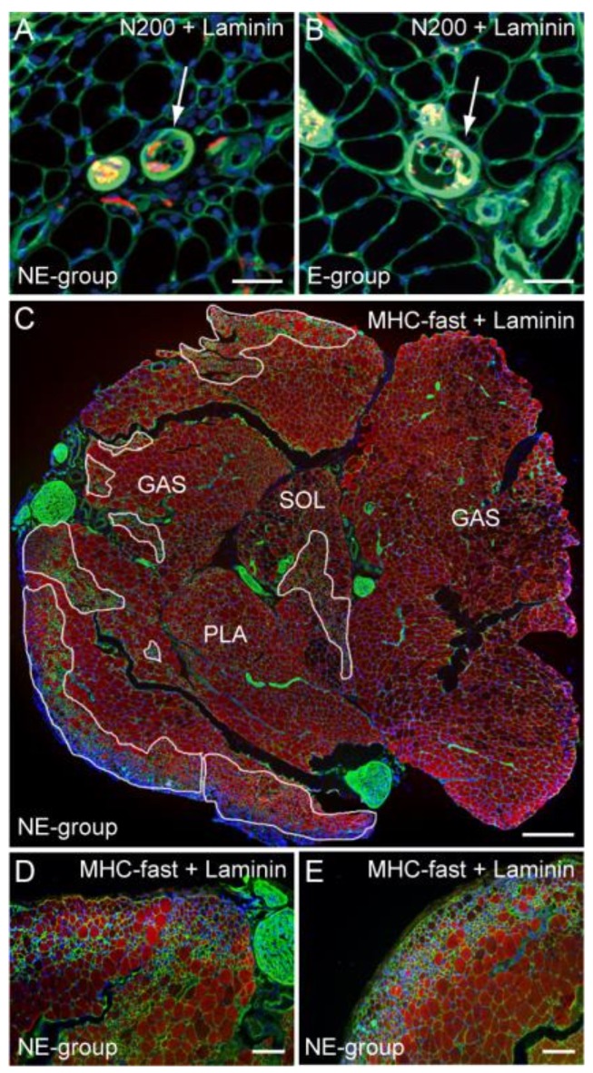
Figure 4.
Detection of muscle spindles and pathological muscle fiber area. Typical muscle spindle in the NE (A, arrow) and E (B, arrow) group stained with N200 (red) and laminin (green). Both spindles are innervated, but irregular fiber diameters and central nuclei are observed in the NE group; (C) Detail of the pathological muscle fiber area in the whole muscle cross-section (GAS, PLA, SOL) stained with anti-myosin heavy chain (MHC) fast type (red) and laminin (green). These enclosed areas account for 17% as pathological area. A higher magnification of the typical pathological muscle fiber features is shown in (D,E). Severe irregular fiber diameters are evident. Blue staining in each panel is the nuclear staining of DAPI. Bars in (A,B) = 50 μm, (C) = 200 μm, and (D,E) = 100 μm.

