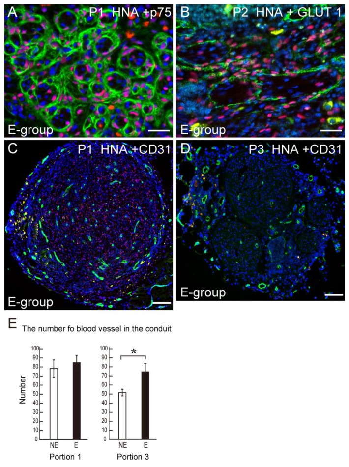
Figure 6.
Differentiation of engrafted human Sk-34 cells into Schwann cells and perineurial cells, and the number of blood vessels in the bridging conduit. (A) Transplanted human Sk-34 cells (pink nuclei) expressed a newly formed Schwann cell marker p75 (green); (B) Transplanted human Sk-34 cells (pink nuclei) also formed new perineurium (green) showing differentiation into perineurial cells; (C) Typical staining of CD31 (green) as evidence of the number of blood vessels in the conduit. Blue staining in each panel is the nuclear staining of DAPI. P1 = position 1, P2 = position 2. Bars in A and B = 50 μm, C = 100 μm; (D) Statistical comparison of the formation of blood vessels in the conduit. There is no difference in the proximal Portion 1 between NE and E group, but a significantly higher value in (E) was observed in the distal end Portion 3. * p < 0.05.

