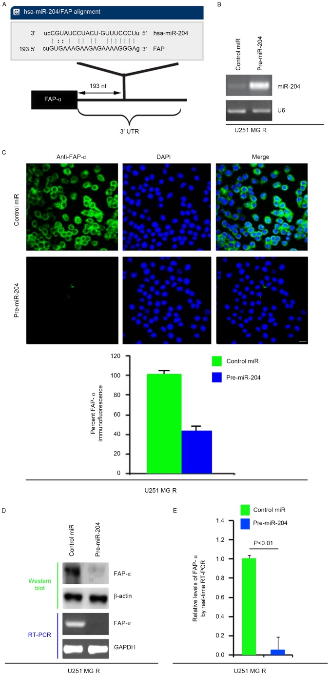
Figure 3.
miR-204 inhibits FAP-α protein expression in U251MG-R cells. (A) Schematic of predicted miR-204 binding sites in the 3′UTR of FAP-α mRNA by miRanda. (B) qPCR for miR-204 in U251MG-R cells. U251MG-R cells were infected with pre-miR-204 or control miR (mock). U6 was the loading control. (n=3). (C) Immunofluorescence analyses for U251MG-R cells transfected with pre-miR-204 and control miR (mock). Upper panel demonstrates microscopy images of immunofluorescence staining of one representative experiment (magnification, ×100). Bottom panel indicates graphic representation of mean fluorescence intensities. (n=3). Scale bar, 20 µm. (D) Western blotting for and RT-PCR for FAP-α protein and FAP-α mRNA, respectively, in U251MG-R cells infected as indicated. β-actin and GAPDH were the loading controls for the western blotting and RT-PCR (n=3). (E) qPCR for FAP-α in U251MG-R cells transfected with pre-miR-204 or control miR (mock). GAPDH was the loading control (n=3). UTR, untranslated region; miR, microRNA; R, resistant; PCR, polymerase chain reaction; RT, reverse transcription; q, quantitative; FAP-α, Fibroblast activation protein α.

