Figure 1.
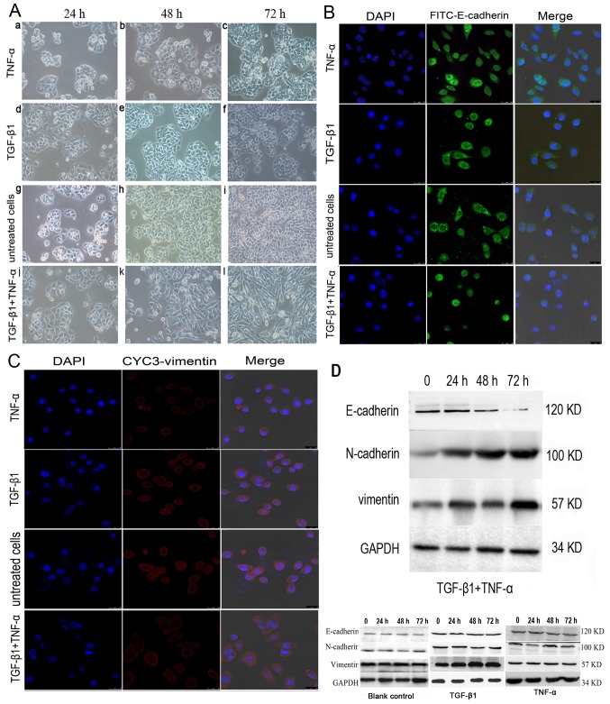
Effects of TGF-β1 and TNF-α on SW480 cells. (A) Morphological characteristics of EMT in SW480 cells treated with (a-c) TGF-β1 or (d-f) TNF-α alone, or in (g-i) combination. (a-f) The majority of TGF-β1 or TNF-α-treated, or (g-i) untreated SW480 cells demonstrated a pebble-like shape and tight cell-cell adhesion at 72 h. (j-l) Cotreatment with TGF-β1 and TNF-α decreased cell-cell contacts and resulted in a more elongated morphological shape compared with individual treatments. (l) TGF-β1 and TNF-α treatment resulted in EMT of SW480 cells, with cells exhibiting a bipolar, spindle-like shape, characteristic of fibroblasts, by 72 h. Magnification, ×200. (B) Immunofluorescent images of SW480 cells treated with TGF-β1, TNF-α or a combination of both for 48 h. Cells were stained with an antibody against E-cadherin (green), and the nucleus was counterstained with DAPI (blue). Subpanels are representative images of the nuclear staining, E-cadherin staining, and a merge of the two, respectively, in SW480 cells. There was a decrease in the an immunostaining of E-cadherin in TGF-β1-and TNF-α-treated SW480 cells compared with cells treated with TNF-α, TGF-β1 alone or untreated cells group. Magnification, ×200. (C) Cells were stained with antibody against vimentin (red), and the nucleus was counterstained with DAPI. Subpanels are representative images of nuclear staining, vimentin staining and a merge of the two, respectively, in SW480 cells. There was an increase in immunostaining of vimentin in TGF-β1- and TNF-α-treated SW480 cells compared with cells treated with TNF-α or TGF-β1 alone, or untreated. Magnification, ×200. (D) Western blotting of the treatment of SW480 cells with TGF-β1 and TNF-α. SW480 cells were incubated with TGF-β1 and TNF-α for 0–72 h and E-cadherin expression was visibly depleted at 72 h. N-cadherin and vimentin expression levels were increased from 24 h. TGF-β1, transforming growth factor-β1; TNF-α, tumor necrosis factor-α; EMT, epithelial-mesenchymal transition.

