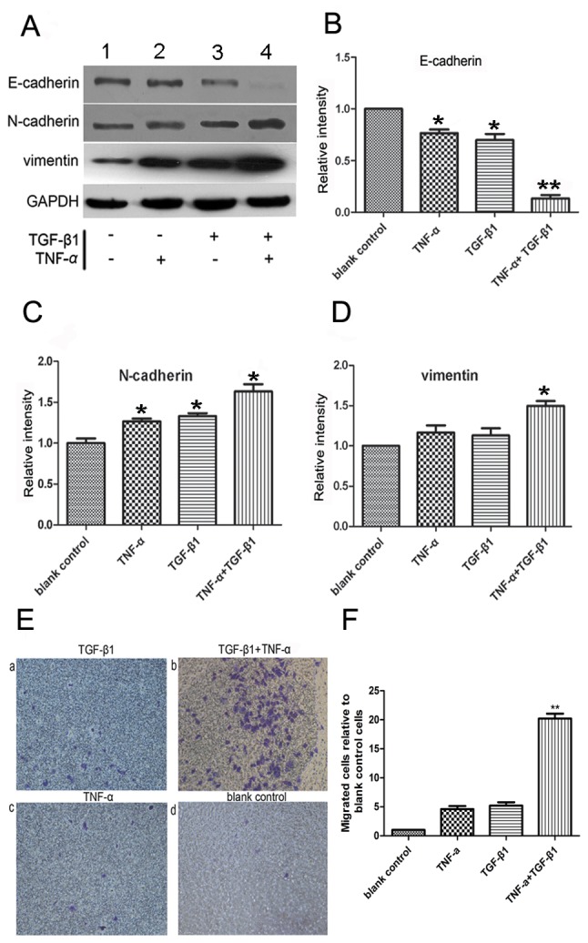
Figure 2.

Analysis of EMT activity of SW480 cells by TGF-β1 and TNF-α. (A) Western blot analysis of the EMT activity of SW480 cells. The relative expression of (B) E-cadherin, (C) N-cadherin and (D) vimentin were examined. TGF-β1 or TNF-α alone decreased E-cadherin expression and increased the expression level of the EMT marker, N-cadherin; *P<0.05 vs. blank control (n=3). Cotreatment with TGF-β1 and TNF-α significantly altered the protein expression levels of E-cadherin, vimentin and N-cadherin; *P<0.05 vs. TGF-β1 or TNF-α alone (n=3). TGF-β1 or TNF-α alone exerted no significant effect on vimentin expression. The results were normalized to the level of GAPDH, used as a loading control. (E) Modulation of the mesenchymal-like properties of SW480 cells by TGF-β1 and TNF-α, (×200 magnification) (a and c) SW480 cells were incubated with TGF-β1 or TNF-α for 72 h, and the number of migratory cells was determined. (b) Cotreatment with TGF-β1 + TNF-α induced an increased number of migratory cells compared with TGF-β1 or TNF-α treatment alone. The majority of cells exhibited a bipolar phenotype characteristic of fibroblasts. (d) Untreated cells demonstrated few or no migrated cells. (F) Cotreatment with TGF-β1 + TNF-α induced an increased number of migratory cells compared with TGF-β1 or TNF-α treatment alone. **P<0.01 vs. TGF-β1 or TNF-α treatment alone (n=3). Values are presented as the mean ± standard error of the mean of triplicate samples. EMT, epithelial-mesenchymal transition; TGF-β1, transforming growth factor-β1; TNF-α, tumor necrosis factor-α.
