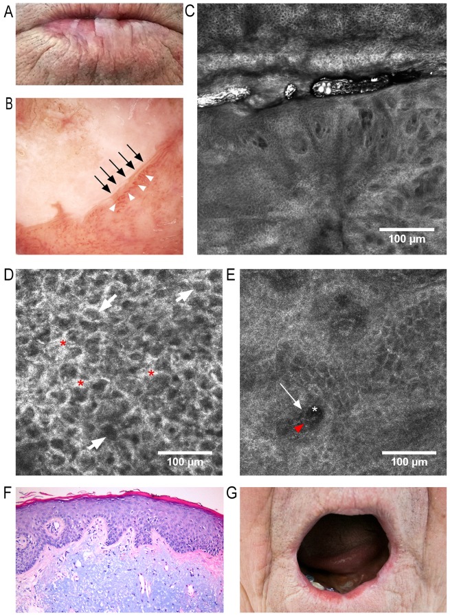
Figure 1.f.
(A) Clinical image: White keratotic areas on the lower lip surface with blurring of skin-vermillion contour. (B) Dermoscopic image: Milky-white plaque with well-defined borders (black arrows) surrounded by dilated and tortuous vessels (white arrowheads). (C) RCM block (dimensions: 2.5×2.5 mm) showing, in the upper third, a keratotic area with impaired keratinocyte architecture, enlarged intercellular spaces and atypical honeycomb appearance, and in the lower two-thirds, dilated blood vessels with tortuous aspect and bright perivascular inflammatory cells. (D) RCM image (500×500 µm) at the stratum spinosum, showing atypical honeycombed pattern, enlarged intercellular spaces (red asterisks), and dark central elements representing nuclei surrounded by bright cytoplasm (white arrows). (E) RCM image (500×500 µm) at the dermo-epidermal junction (DEJ)/papillary dermis, revealing dark areas representing papillary blood vessels (white asterisk) containing central elements corresponding to blood cells (red arrowhead) and bright perivascular structures equivalent to perivascular inflammatory cells (white arrow). (F) Histopathological image (hematoxylin and eosin staining; magnification, ×200) of AC, showing hyperkeratosis and irregular acanthosis, mildly atypical keratinocytes within the lower third of epithelium, and marked solar elastosis and vascular ectasia within the dermis. (G) Clinical image captured 7 months after classic vermilionectomy, with minimal scarring and deformity, and with no signs of recurrence. RCM, reflectance confocal microscopy.

