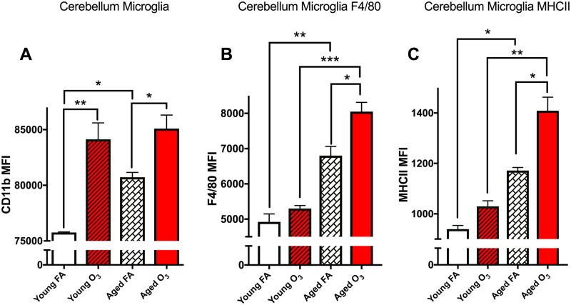
Figure 3.
Acute O3 exposure increases primed microglia in the aged cerebellum. The microglia population was further analyzed for CD11b, F4/80, and MHCII expression levels, as measured indicative of their “activation” phenotype, or reactive gliosis. A, Bar graph illustrating the CD11b mean fluorescence intensity (MFI) of microglia in the cerebellum. Age significantly increases this population and acute O3 exposure increases this population in both young and aged cerebellar tissue. B, Bar graph illustrating the F4/80 MFI of the microglia population indicative of reactive gliosis or “priming” of microglia in the cerebellum. Age significantly increases this population, and acute O3 exposure increases this population only in the aged cerebellum. F4/80 expression in the aged cerebellum after acute O3 exposure is significantly higher than in the young cerebellum after exposure. C, Bar graph illustrating the MHCII MFI of the microglia population indicative of reactive gliosis or “priming” of microglia in the cerebellum. Age significantly increases this population, and acute O3 exposure increases this population only in the aged cerebellum. MHCII expression in the aged cerebellum after acute O3 exposure is significantly higher than in the young cerebellum after exposure (n = 3 per group, *p < .05; **p < .01; ***p < .001; ****p < .0001 by 2-way ANOVA; all values represent mean ± SEM).

