Figure 3.
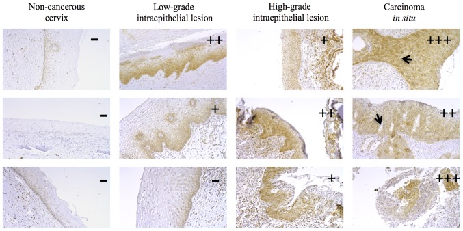
KCNMA1 protein expression in human cervical dysplasia and carcinoma. The images show variable immunostaining intensity of KCNMA1 protein expression (brown immunostaining). Non-cancerous human biopsies were negative for KCNMA1 immunostaining. Low-grade cervical dysplasia samples displayed the lowest intensity, while the stronger immunostaining was observed in the carcinoma in situ biopsies. The staining intensity was scored as indicated under Materials and methods. The arrows indicate the rupture of the basal membrane, probably caused by the proliferation of cervical cancer cells. Representative images from six independent experiments in each condition are shown. Magnification, ×20.

