
Figure 3. Electrophysiological characterization of TRPML1 and its agonist-binding pocket.
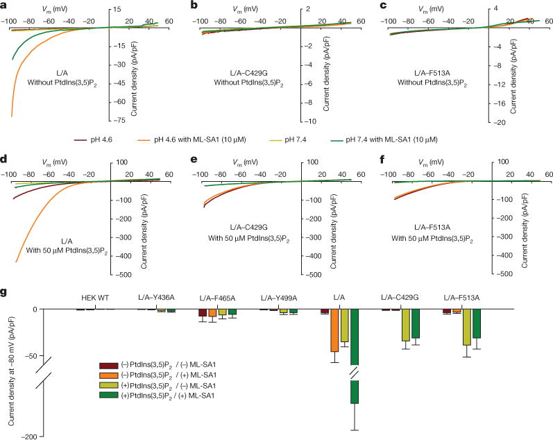
a, Whole-cell currents of HEK293T cells transfected with surface-expressing eGFP-TRPML1 with leucine-to-alanine mutants (L15L and L577L to alanine; abbreviated L/A), and L/A with binding pocket mutations. b, c, C429G (b) and F513A (c) with or without 10 μM ML-SA1 at pH 4.6 or pH 7.4 without PtdIns(3,5)P2. d–f, Whole-cell currents for constructs from panels a–c with 50 μM PtdIns(3,5)P2 substituted in the cytoplasmic solution. g, Whole-cell current density at −80 mV recorded at pH 4.6 for cells transfected with empty vector (wild type, WT), L/A, and L/A plus Y436A, F465A, Y499, C429G or F513A individually. Values are mean ±s.e.m.