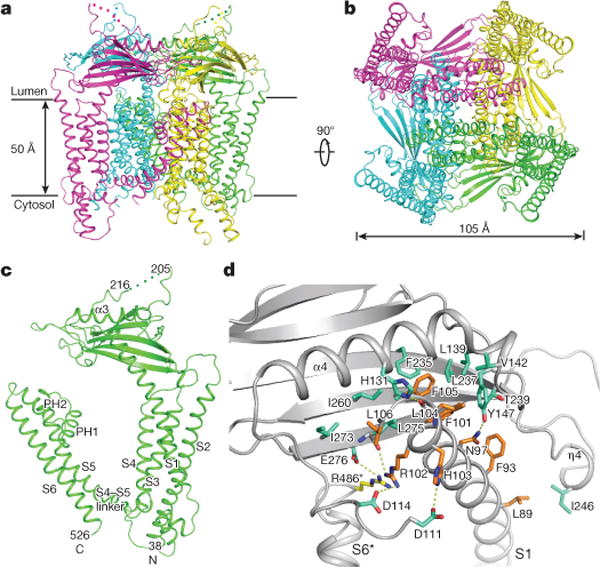
Figure 1. Overall structure of TRPML1.

a, Ribbon representation of the structure in perspective horizontal to the plane of the membrane, with four subunits coloured differently. The flexible linker in the lumenal domain is indicated by dots. b, Structure rotated 90° around a horizontal axis. c, Ribbon representation of a TRPML1 subunit with different domains denoted. d, The interface between a transmembrane region and the luminal domain. Luminal domain residues are coloured green, S1 residues are orange, and the arginine residue from the neighbouring unit is yellow. All hydrophilic interactions are indicated by dotted lines. S6* denotes S6 of the neighbouring subunit.
