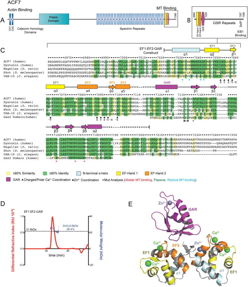
Figure 1. Architecture of the ACF7 EF1-EF2-GAR Module.
(A) hACF7 domain architecture and zoom view (B) of the MT-binding region. (C) Sequence alignment of the EF1-EF2-GAR module from human, D. rerio, D. melanogaster, and C. elegans spectraplakins, and human Gas2. hACF7 EF1-EF2-GAR 2° structure and residue number are depicted above, residues mutated are indicated below. (D) SEC-MALS analysis of the EF1-EF2-GAR module. The molecular weight of a monomer is indicated by the dashed line. The peak measured accounts for 96.4% of the total mass eluted. (E) Model of the EF1-EF2-GAR modules observed in the ASU. See also Figure S1.

