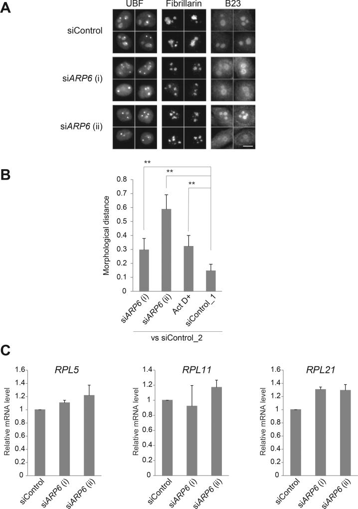
Fig. 3.
Morphological changes of the nucleolus upon ARP6 depletion. (A) HeLa cells were transfected with siRNA targeting ARP6 and immunostained with antibodies against UBF, fibrillarin, and B23, specific markers for the nucleolar subcompartments FC, DFC, and GC, respectively. Four representative images for each staining are shown. Scale bar, 10 µm. (B) Quantification of morphological difference of the UBF-stained nucleoli was performed with wndchrm, to compute degree of morphological distance from siControl_2. A large value of the morphological distance indicates that the morphology of the cells is different from the morphology of siControl_2 cells. siControl_1 was used as a negative control. Image sets used in this analysis are shown in Supplementary Fig. 2. Values are means ± standard error of 20 cross validation tests; **, p < 0.01. (C) Ribosomal protein gene expressions in ARP6 knockdown cells. The mRNA was measured with RT-qPCR. GAPDH mRNA expression was used as the internal control. Expression levels of control cells are set to 1. Values are represented as means ± standard error of triplicates.

