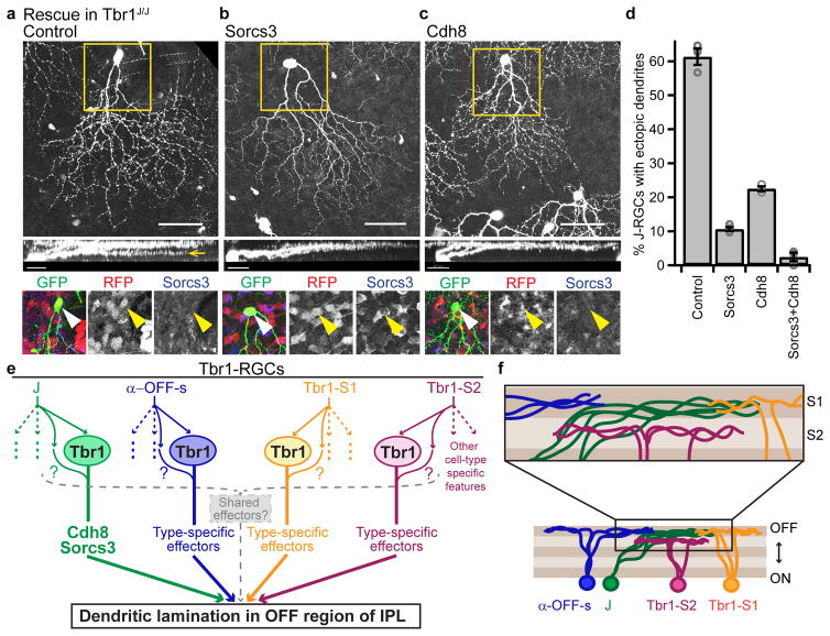
Figure 8. Regulation of dendrite laminar identity by Tbr1.
(a–c) En face (scale=50μm) and rotated (scale=25μm) views of Tbr1 mutant J-RGCs that had been infected with a control AAV (a), a Sorcs3-expressing AAV (b) or a Cdh8-expressing AAV (c). Sorcs3 was co-injected with RFP while Cdh8 was RFP-tagged. Yellow boxes mark insets, which show expression of AAVs and Sorcs3 protein in J-RGCs. As expected, Sorcs3 protein is lost from Tbr1 mutant J-RGCs infected with control or Cdh8 AAVs; it is restored in Sorcs3-infected J-RGCs (yellow arrowheads). Dendritic defects in Tbr1-deficient J-RGCs (a, yellow arrow) were rescued by either Sorcs3 (b) or Cdh8 (c). (d) Proportions of J-RGCs with ectopic dendrites in Tbr1J/J retinas. n = 3 control, 4 Sorcs3, 3 Cdh8 and 3 Sorcs3+Cdh8 infected animals. p<0.0001, F(3,9)=376, one-way ANOVA; p<0.0001 for control versus Sorcs3, Cdh8 or Sorcs3+Cdh8. Tukey-Kramer test. (e) Model of Tbr1-regulated laminar patterning. Solid, colored arrows indicate molecular pathways that specify lamination. Dotted, colored arrows indicate pathways for other cellular features. Arrows emanating from ovals marked ‘Tbr1’ indicate cell-type specific effectors that bring dendrites to the OFF strata of the IPL, such as Cdh8 and Sorcs3 in the case of J-RGCs. Colored question marks post the hypothesis that Tbr1-independent mechanisms may act in parallel to Tbr1 to specify lamination. Dotted grey bracket and arrow indicate the possible presence of shared effectors for dendritic lamination that are commonly expressed by all four types. (f) IPL schematic showing that all four Tbr1-RGC types laminate within the OFF half of the IPL (bottom), but differ in their precise lamination.

