
Figure 1. Adipocytes induce CD36 expression in ovarian cancer cells.
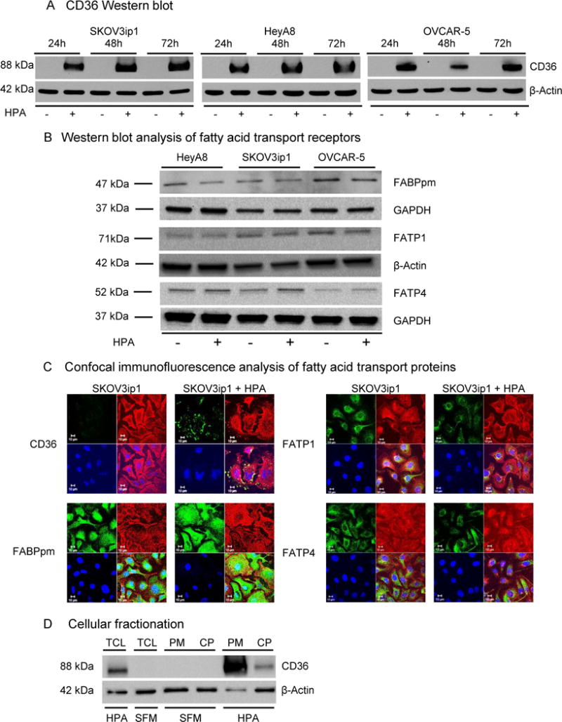
(a) Western blot analysis of CD36 protein expression in OvCa cell lines co-cultured with human primary adipocytes (HPA) at the indicated time points.
(b) Western blot analysis of FA transport proteins FABPpm, FATP1 and FATP4 in three different OvCa cell lines co-cultured with HPA for 24 h.
(c) Confocal immunofluorescent microscopy for the indicated fatty acid transporters in the absence (-) and presence (+) of HPA. Fatty acid transporters are detected using green fluorescence, plasma membrane using red fluorescence (Wheat Germ Agglutinin), cell nuclei are counterstained with Hoechst 33258 (blue).
(d) Subcellular localization of CD36 protein. OvCa cells were either co-cultured with HPA or were grown in serum free media (SFM). After 16 h, cells were either lysed to obtain total cellular lysate (TCL) or fractionated to cytoplasmic (CP) and plasma membrane (PM) fractions.