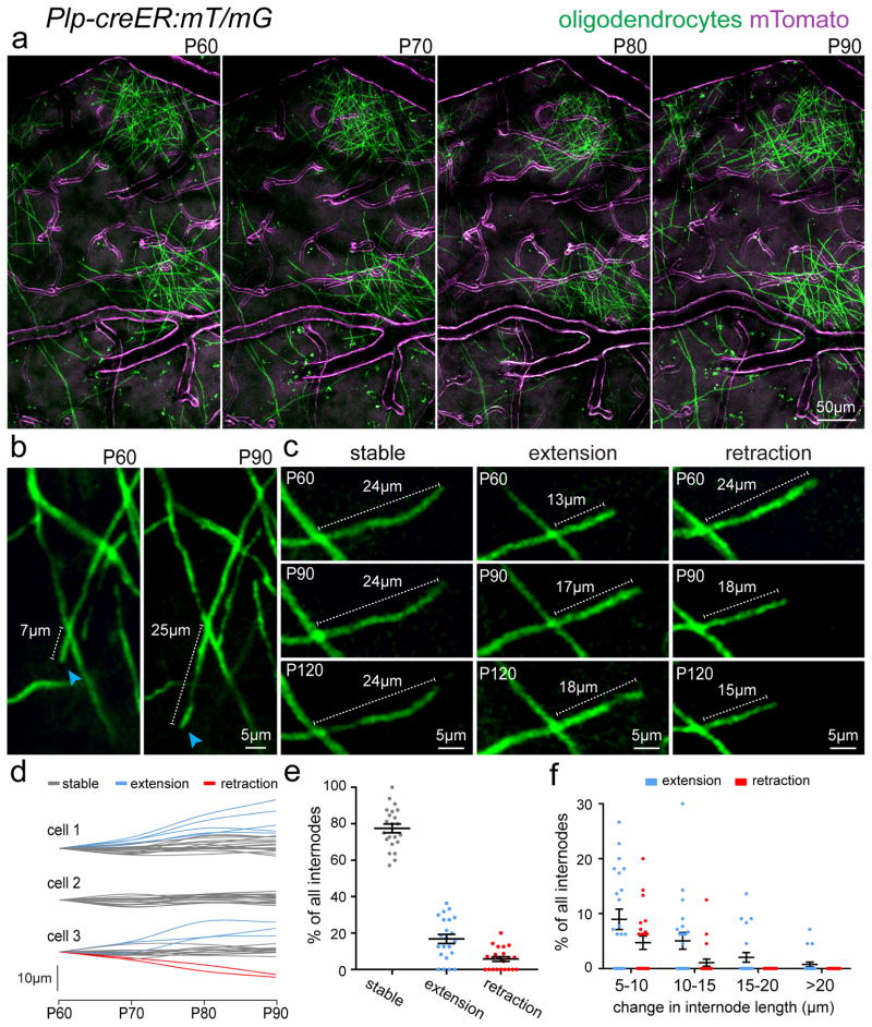
Figure 4. Evidence of myelin plasticity through internode remodeling.
(a) In vivo two-photon fluorescence images of single oligodendrocytes imaged over 30 days in a transgenic mouse line (Plp-creER:mT/mG) with membrane tethered GFP (mGFP) expressed specifically in mature oligodendrocytes and membrane tethered Tomato (mTomato) expressed predominantly in cerebral blood vessels. (b) In vivo time-lapse images showing extension of a single internode (blue arrowheads) with stability of all other internodes in the field of view. (c) In vivo time-lapse images showing the three observed behaviors for internode length changes over time. (d) Measurements of changes in single internode length for individual oligodendrocytes as indicated. Single cells exhibited heterogeneous internode plasticity over the 30 days of imaging with single internodes remaining stable (gray lines), extending (blue lines), or retracting (red lines). (e–f) Distribution of the single internode behavior in all cells. The vast majority of internodes showed no change (>5μm) in length however a proportion displayed long-term extension or retraction over the imaging period ranging from 5–20μm changes over 30 days of imaging as indicated, data represent 22 individual oligodendrocytes, 330 internodes, from 3 mice. Dots indicate single oligodendrocytes and error bars are mean +/− s.e.m., each image is representative of at least three locations in at least three animals.

