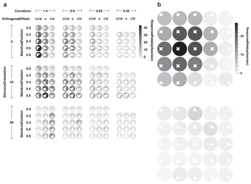
Fig. 1. Transformation of retinotopic information into contour coding in area V4.
This neuron exemplifies how precise retinotopic information is recoded in terms of contour orientation, curvature, and object-relative position. The stimuli shown here (white shapes) were derived from a more wide-ranging test of shape sensitivity that revealed tuning for sharp convex curvature pointing (oriented) toward the upper right and positioned to the upper right of object center. (a) This fine-grained test shows gradual tuning for curvature (horizontal axis), orientation (vertical axis), and object-relative position (recursively plotted within axes) of the convex curvature. Response rate for each shape is indicated by background shade (see scale bar at right). (b) This test demonstrates how a shape with the critical convex curvature at its top right drives responses across a broad range of retinotopic positions (top), while a similar shape without this feature elicits little response (bottom). Adapted from Pasupathy and Connor (2001)27.

