
Figure 2. Example subfield boundaries from one individual monkey.
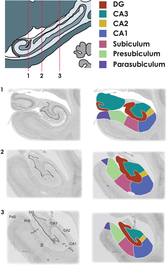
Subfield boundaries drawn at three anterior-posterior locations along the extent of the hippocampus (1-most anterior, 3-most posterior). Once a subject’s boundaries are completed, each subfield is assigned a unique identifying color. Multicolor mask images are then converted to separate binary images for each hippocampal subfield for further processing.
[pml - polymorphic layer, gl - granule cell layer, slm - stratum lacunosum-moleculare, sr – stratum radiatum, pcl - pyramidal cell layer, so - stratum oriens, al – alveus, ilf - inferior longitudinal fasciculus]