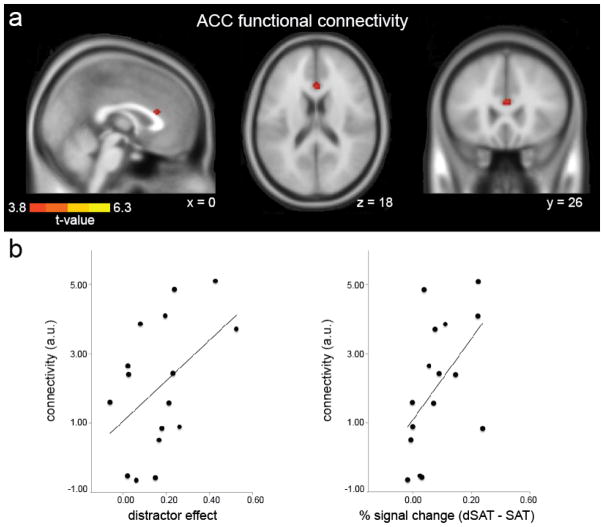
Figure 4. PPI functional connectivity during distractor challenge.
(a) Psychophysiological interaction (PPI) analyses revealed greater functional connectivity between the right PFC (RPFC) seed region (8 mm sphere centered on IFG peak coordinates MNI 46, 2, 30) and anterior cingulate cortex (ACC) during distractor challenge. RPFC also showed increased connectivity with regions listed in Table 3, medial frontal gyrus/supplementary motor area and superior temporal gyrus (not displayed). T-maps are displayed on an SPM template average of 152 normalized T1 anatomical scans, p < .001, k > 20 (see Methods). (b) Increased right ACC - RPFC functional connectivity (arbitrary units, a.u.) was associated with greater performance declines during distractor challenge and greater increases in right RPFC activation. Functional connectivity strength showed a modest relationship between the distractor effect on performance (SAT – dSAT score), r = .48, p = .07, and increased RPFC activation, r = .55, p = .03.

