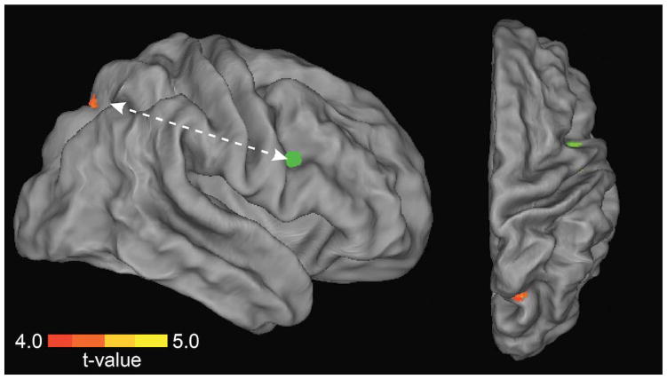
Figure 5. Frontoparietal functional connectivity associated with preserved performance during distractor challenge.
Multivariate regression analyses identified a region in right precuneus/superior parietal lobule (SPL, warm colors) whose functional connectivity with right PFC (8 mm sphere centered on inferior frontal gyrus peak coordinates MNI 46, 2, 30) was greatest for individuals with low behavioral impact of distraction. Green indicates the location of the seed.

