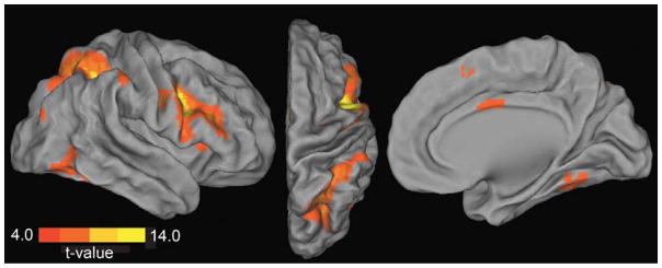
Figure 6. Resting state functional connectivity before task performance.
Regions showing positive synchronization with the right PFC seed region (8 mm sphere centered on inferior frontal gyrus peak coordinates MNI 46, 2, 30) during the resting state scan collected prior to task performance are displayed. Activity in right PFC was correlated with other task positive regions including superior and inferior parietal cortex, middle frontal gyrus, inferior temporal gyrus (lateral and dorsal views), and cingulate cortex (medial view). Displayed activations are at p < .001, k > 20; see Table 3 for FDR correction.

