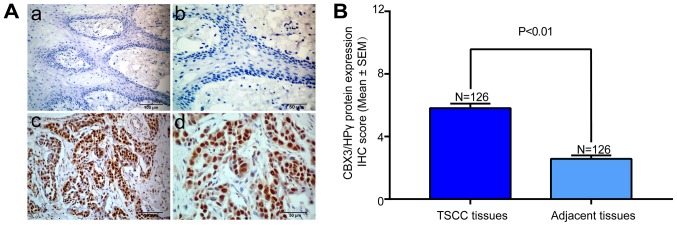
Figure 2.
Immunohistochemical staining of CBX3/HP1γ protein in TSCC tissues and cancer-adjacent normal tissue. (Aa) Representative weak staining of CBX3/HP1γ (low expression) in cancer-adjacent normal tissue (magnification, ×200). Nuclei were counterstained with hematoxylin. (Ab) Magnified image from a (magnification, ×400). (Ac) Representative strong staining of CBX3/HP1γ (high expression) in primary TSCC sample (magnification, ×200). (Ad) Magnified image from c (magnification, ×400). CBX3/HP1γ expression was identified primarily in nuclei of cancer cells. (B) Statistical analysis of CBX3/HP1γ IHC scores in TSCC tissues and cancer-adjacent normal tissue. Data was presented as the mean with SEM.

