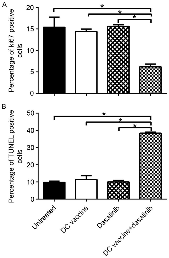
Figure 3.

Changes in the proliferation and apoptosis of 4T1 tumor cells after different treatments. Immunohistochemical analysis of Ki67 in each group of tumors. (A) Ki67-positive cells in each group of tumors were measured. Immunohistochemical analysis of apoptosis in each cohort of tumors. (B) TUNEL positive cells in each group of tumors were measured. *P<0.05. DC, dendritic cell; TUNEL, terminal deoxynucleotidyl transferase dUTP nick end labeling.
