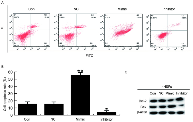
Figure 4.
Effects of miR-26a on hHSF cell apoptosis. (A and B) At 48 h post-transfection, flow cytometry was used to assess the effect of miR-26a on hHSF cell apoptosis. (C) Effects of miR-26a on the protein expression levels of Bcl-2 and Bax were analyzed by western blotting. Data are presented as the mean ± standard deviation. *P<0.05 and **P<0.01 vs. Con. miR, microRNA; hHSFs, human hypertrophic scar fibroblasts; Bcl-2, B-cell lymphoma 2; Bax, Bcl-2-associated X protein; Con, control group, cells without any treatment; FITC, fluorescein isothiocyanate; inhibitor, cells transfected with miR-26a inhibitor; mimic, cells transfected with miR-26a mimic; NC, negative control group, cells transfected with the negative control vector; PI, propidium iodide.

