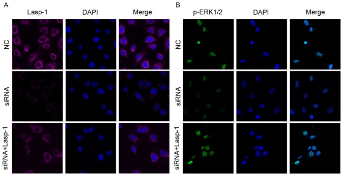
Figure 3.
Lasp-1 and p-ERK1/2 protein expression following Lasp-1 knockdown. (A) Lasp-1 protein expression detected using immunofluorescence. The Merge panels are superimposed images of Lasp-1 staining in red and nuclei staining in blue (using DAPI). Magnification, ×400. (B) p-ERK1/2 protein expression detected using immunofluorescence. The Merge panels are superimposed images of p-ERK1/2 staining in green and nuclei staining in blue (using DAPI). Magnification, ×400. Lasp-1, LIM and SH3 protein 1; ERK, extracellular-signal-regulated kinase; p-, phospho-; NC, negative control; siRNA, small interfering RNA.

