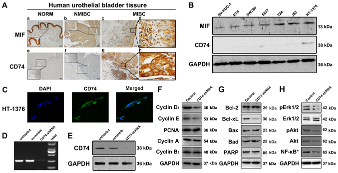
Figure 1.
Expression of MIF and CD74 in tissue samples and cells, and protein expression levels in HT-1376 cells following knockdown of CD74. (A) Little or no expression of immunoreactive CD74 was identified in the urothelial layers of normal bladder and NMIBC samples, but sections of MIBC samples exhibited strong immunoreactive signals of CD74. In contrast, MIF demonstrated strong immunoreactions in the normal and UCB samples. (B) Western blotting assay results for MIF and CD74 in the cultured urothelial SV-HUV-1, SW780, 5637, T24, J82 and HT-1376 cell lines. HT-1376 was the only one identified to express CD74, but all cells expressed MIF to a certain extent. (C) Immunofluorescence microscopy (×100, magnification) indicated positive CD74 staining in HT-1376 cells. (D) CD74 shRNA lentiviral particles abrogated the RNA expression of CD74 in the HT-1376 cells compared with untreated and scramble cells. (E) CD74 short hairpin RNA lentiviral particles abrogated the protein expression of CD74 in the HT-1376 cells when compared with the untreated and scramble cells. (F) Knockdown of CD74 modulated the expression levels of not only Cyclin D1, Cyclin E, but also intranuclear NF-κB p65, pAkt, pErk1/2. (G and H) However, no significant changes in Bcl-2, Bad, cleaved PARP and Erk1/2 were observed among all groups. *intranuclear NF-κB. NMIBC, non-muscle-invasive bladder cancer; MIBC, muscle-invasive bladder cancer; UCB, urothelial cell carcinoma of the bladder; MIF, macrophage migration inhibitory factor; CD74, cluster of differentiation; p65, transcription factor p65; Akt, RAC-alpha serine/threonine protein kinase; p, phosphorylated; Erk1/2, Extracellular regulated protein kinase 1/2; Bcl-2, B-cell lymphoma 2; Bcl-xL, Bcl-extra large; Bax, Bcl-2-associated X protein; PARP, poly(adenosine 5′-diphosphate-ribose) polymerase.

