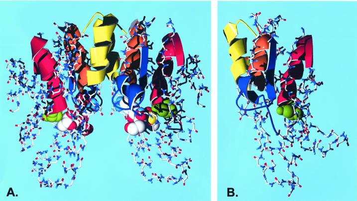
Figure 6.
Three-dimensional structure of plant GSTs with the strongly conserved type III features mapped. The active site Ser is shown in green as a space-filled model. The conserved patches in the Type III consensus sequence are shown as ribbons and colored as red, S20-L38; blue, K49-H68; orange, E76-E86; and yellow, L101-W114. A, The lactoylglutathione complex of a GSTI dimer taken from Neuefeind et al. (1997a). The substrate analog is shown as a space-filled model using Corey, Pauling, and Koltun colors. The regions of GSTI that are homologous to the type III conserved patches are S11-E29 (red), K41-N58 (blue), E66-R76 (orange), and R84-W98 (yellow). B, A homology model of ZmGST 24 prepared as described in the text. The monomer is shown in the same orientation as the GSTI dimer. The conserved patches in the ZmGST 24 sequence are S11-E29 (red), K38-H57 (blue), E64-E74 (orange), and L85-W98 (yellow).

